Comparison between autosegmentation and clinical contours for whole heart and associated differences in whole heart dosimetry metrics
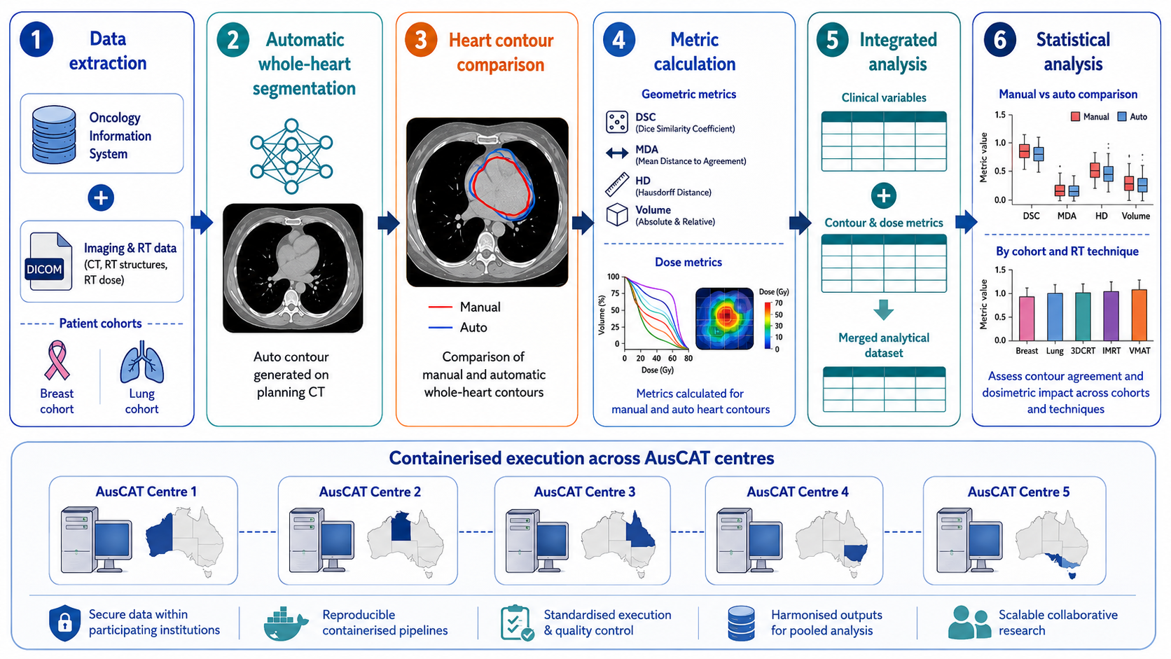
Radiation therapy is a common treatment for breast and lung cancer, but it can sometimes expose the heart to radiation, which may increase the risk of long-term heart diseases. Clinicians use measurements such as the average dose of radiation the heart receives to estimate this risk. While the dose itself is usually calculated consistently across hospitals, there is uncertainty in how clinicians contour the heart on the scans used for planning the treatment. These contours are important because they define which part of the scan is considered the heart, and even small differences may change the calculated heart dose.
In this project, we are investigating three main questions:
-
How consistent are heart contours between patients and across different hospitals?
-
Do variations in contouring the heart affect the calculated dose the heart is thought to receive?
-
If dose estimates vary, could this change the predicted risk of heart side effects from radiation?
To answer these, we are comparing the contours drawn by clinicians with those generated by machine learning-based auto-segmentation tools. We will also study how these differences impact heart dose calculations and whether they could influence models that predict long-term heart risks.This research will help us understand whether using automated contouring tools can improve consistency in measuring heart radiation exposure and ultimately lead to better protection of heart health for people undergoing radiotherapy.