Understanding Radiation Exposure to Heart substructures in Cancer Radiotherapy
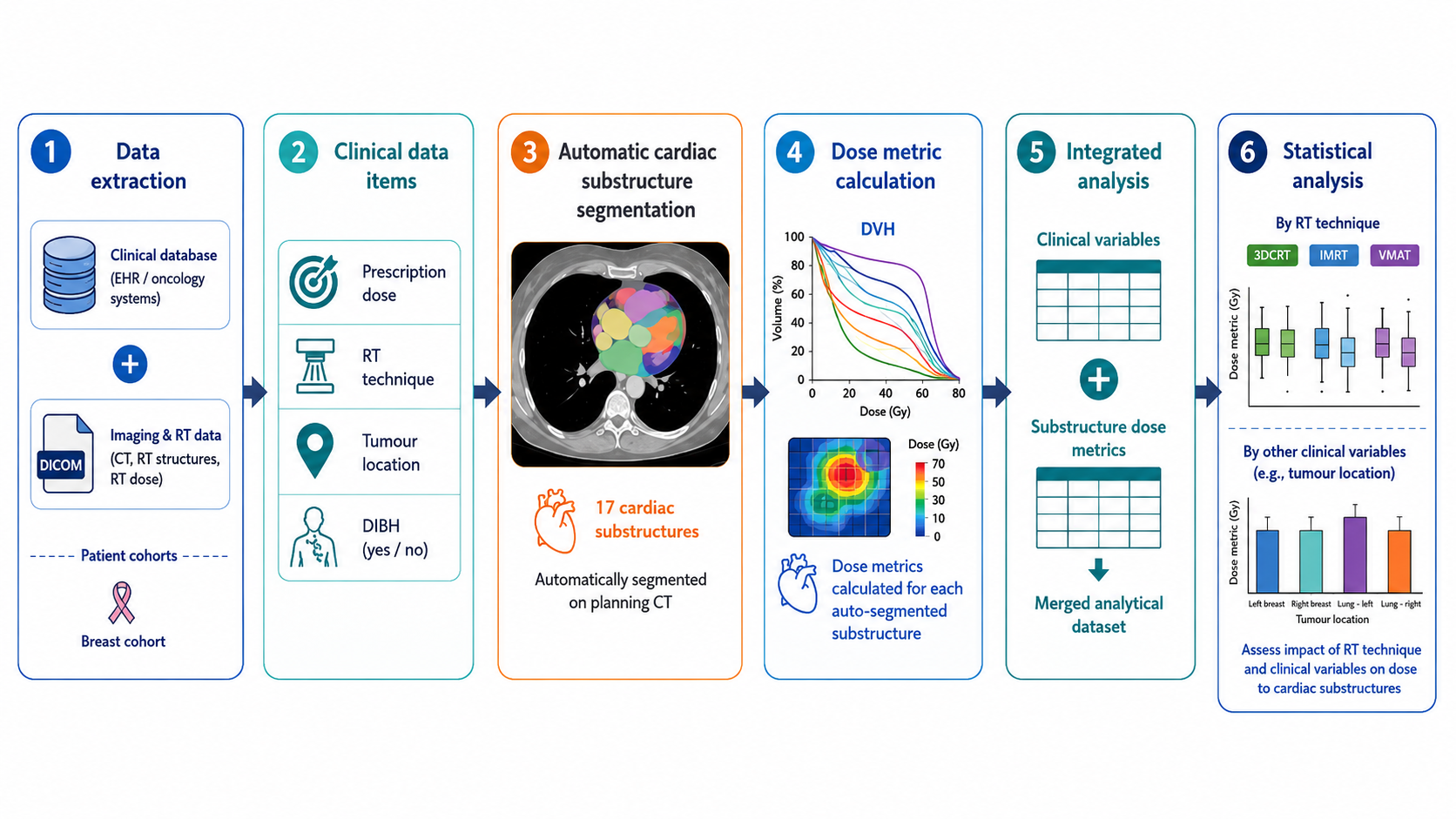
Radiation therapy is commonly used to treat breast and lung cancer patients, but one of its potential side effects is damage to the heart. Traditionally, studies have focused on the average radiation dose to the whole heart when assessing this risk. However, new research suggests that the heart (such as Pulmonary artery or Left ventricle) may also be sensitive to radiation, and damage to these areas could increase the risk of heart diseases, even if the overall heart dose appears safe.
The challenge is that manually contouring these small heart regions is very time-consuming, so past studies have been limited to only a few substructures and relatively small patient groups. Our team has developed an open-source tool that can automatically contour the whole heart and 17 individual cardiac substructures. Using this tool, we can now analyse radiation doses to the heart in much larger numbers of breast and lung cancer patients treated across multiple hospitals.
This study aims to answer:
-
What levels of radiation do different heart substructures receive in breast and lung cancer treatment?
-
How do different treatment techniques (such as 3D conformal therapy, IMRT, and VMAT) affect these doses?
-
Do other factors, like where the tumour is located, change how much radiation reaches different parts of the heart?
By better understanding which parts of the heart are most exposed to radiation, this research will help guide safer treatment planning and reduce the risk of long-term heart complications for cancer patients.